60 Strathallen Avenue, NORTHBRIDGE

Opening Hours : Monday to Friday 9am to 5pm
  Contact : 02 9958 0552

All Posts in Category: case studies

Case Studies: Left Branch Retinal Vein Occlusion


Case Studies: Left Branch Retinal Vein Occlusion

A 40 year old patient presented with sudden onset of painless vision loss from his left eye. There were no associated ocular symptoms and he was systemically well, though moderately over weight.

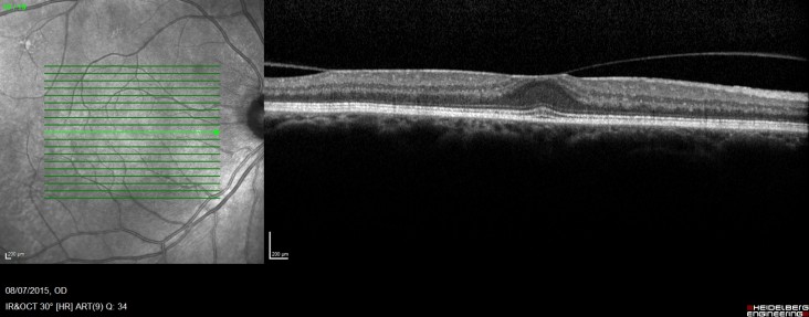
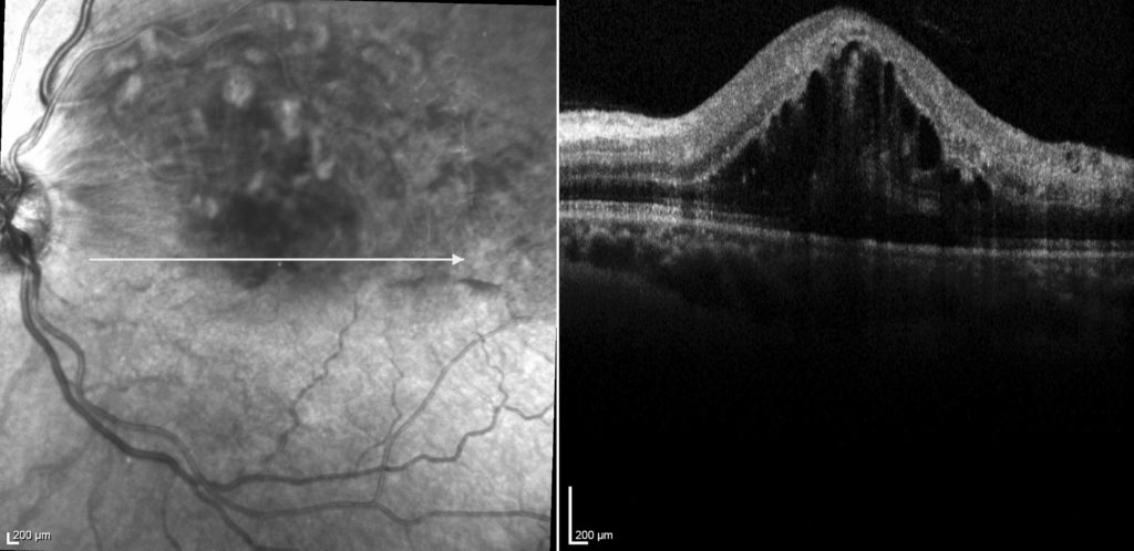
On examination his corrected  acuities were right 6/6 and left 6/90. His intraocular pressures were normal. His anterior segments were unremarkable with clear ocular media and no rubeosis. Dilated fundus examination on the right was essentially unremarkable and on the left there were  retinal haemorrhages and cotton-wool spots, along the superotemporal arcade, consistent with a branch retinal vein occlusion. On OCT scanning, he had significant associated macular oedema (see figure 1).
NADER, Michael, 1859

FIG 1

The patient was diagnosed with a left branch retinal vein occlusion. The most common cause underlying  cause is atherosclerotic disease, associated with systemic hypertension. However, given this patient was only 40 years of age, obstructive sleep apnoea and pro-coagulant diseases need to be ruled out.

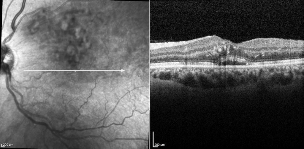
Treatment of intravitreal Lucentis injection therapy was instituted, and four weeks after the first injection his vision returned to 6/9 and his macular oedema dramatically improved, as seen in figure 2 below.
NADER, Michael, 1859

FIG 2

Read More

Case studies: Bilateral Proliferative Diabetic Retinopathy

I saw a patient, who presented to his optometrist initially due to a floater in his left eye.

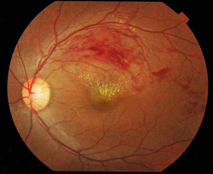
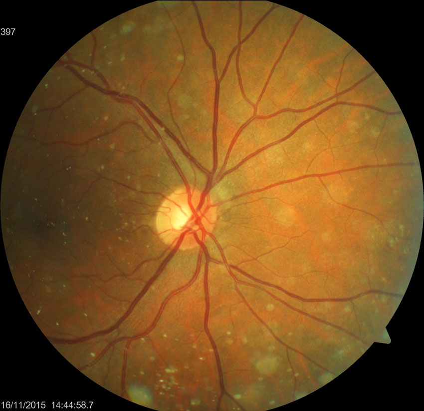
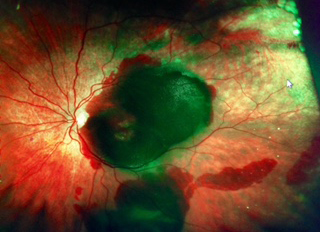
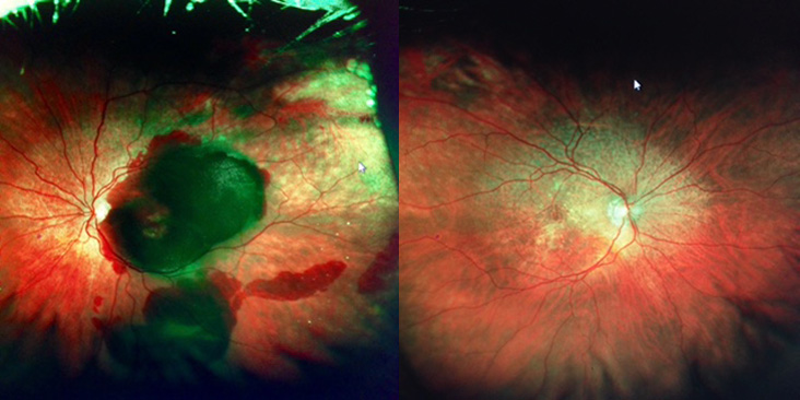
On examination today, his unaided acuities are right 6/12+ and left 6/6, the right improved to 6/9- with a pinhole. His intraocular pressures were right 16mmHg and left 14mmHg with Goldman applanation tonometry. Dilated fundus examination revealed bilateral proliferative diabetic retinopathy. He has pre-retinal haemorrhages on the right. See pictures. There were no signs of retinal tears

This patient has bilateral proliferative diabetic retinopathy. Given he did not have a diagnosis of diabetes, we checked his sugar level, which was 20mmol/L. He has been sent to his GP for systemic assessment. Meanwhile a combination treatment of intravitreal injections and retinal laser has been advised for his eyes.

Read More

Case studies: Synchysis Scintillas

Synchysis-scintillans2

A 61 year old female presents for a review of drusen in her right eye.

With glasses her visual acuity is right 6/12 improving to 6/7.5 with a pinhole, and left 6/9, improving to 6/7.5. Her intraocular pressures were 14mmHg bilaterally. Retinal examination revealed no drusen however we found Synchysis Scintillans in her right eye.

Synchysis scintillans is a vitreous disorder that is usually compared with asteroid bodies in the vitreous. These are extremely rare cholesterol crystals that move around freely in the vitreous and falls to the floor of the vitreous due to gravity. No treatment is required.

Read More

Case studies: Macroaneurysm

I saw a patient, who had right sided cataract surgery under my supervision at Royal Prince Alfred Hospital earlier this month.

He presented today with unaided acuities of right 6/9+ and left 6/12. Everything appeared as planned with normal intraocular pressures, clear corneas and a well centred intraocular lens. Dilated fundus examination however, revealed an intra-retinal and pre-retinal haemorrhage along the infero-temporal vascular arcade, consistent with a macroaneurysm. Thankfully his fovea and central macula is unaffected.

In this patient’s case, I will send him back to his GP for monitoring of his blood pressure, as hypertension is the most common risk factor here. Review is planned for 3 weeks’ time.

Read More

Case studies: Optic Nerve Pit with sub retinal fluid

Optic Nerve Pit OCT

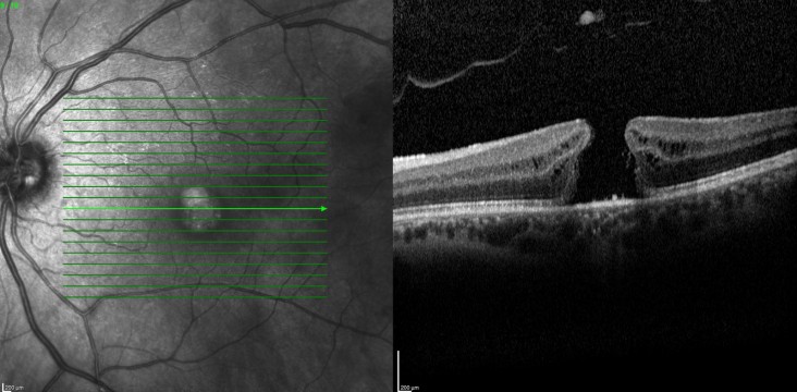
This is a photo of a 17 year old female I saw last week. This is an Optic Nerve Pit with sub retinal fluid. It is a congenital condition. She presented with a 3 day history of blur and distortion in the right eye, with the blurring and distortion moving to the central part of her visual field 1 day ago. She presents acuities of the right 6/90, no improvement with pinhole and the left 6/6.

Normally, an isolated optic nerve pit would not call for any treatment. However, due to the sub-retinal fluid, this patient will require a vitrectomy. I’ll keep you updated on his progress.

Read More

Case studies: Vitreomacular Traction

This is a photo of a 65 year old male I saw last week. This is vitreomacular traction however, he presents with excellent vision – 6/4.5 bilaterally.

At this stage he doesn’t require treatment, but he has been advised to return in a few months. Meanwhile, he will self-monitor with an Amsler Grid and knows to come back if his vision changes or he notes any distortion. I’ll keep you updated on his progress.

Read More

Case studies: Emphysema


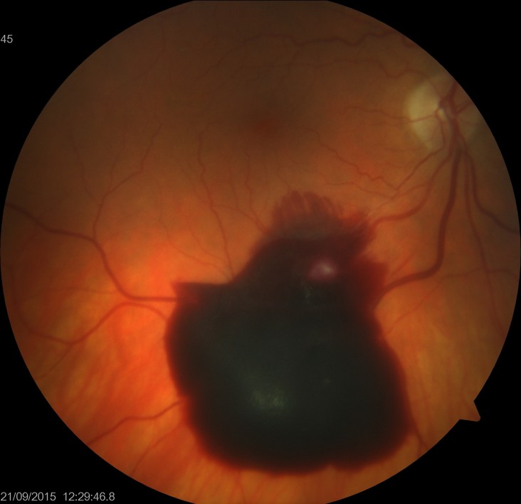
EmphysemaThis is a photo of a lady I saw yesterday. She is 82 years old and suffers from emphysema. Despite this, she still smokes. She has had cataract surgery in the past and presented with a four day history of decreased left vision.

On examination, her visual acuity was Right 6/12 and left Count fingers only.

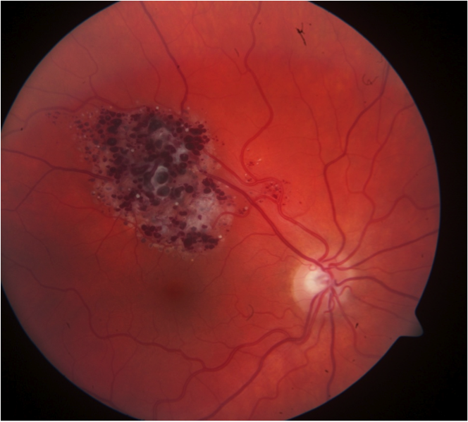
As you can see, she has atrophic changes at her right macula and a huge left macular hemorrhage. I made a diagnosis of wet AMS and injected Lucentis intravitrally.

I asked her to use Macuvision tablets and stressed that she must stop smoking. Let’s see how she goes, I’ll keep you updated.

Read More